Eye Conditions

At Lake Austin Eye, we know that you have questions about eyes diseases and disorders.

Will I get cataracts? What is glaucoma, and what are the symptoms and treatment options? How long does it take to recover from macular degeneration treatment? We hope the following FAQ provides a better understanding for you on eye disorders and your treatment options.

Eye Conditions FAQ

  • What are Cataracts?

    About 50% of all Americans ages 65 to 75 have cataracts to some degree. Cataracts occur normally as we age, causing our natural lens to become cloudy. They obstruct the passage of light, causing impaired vision. Since cataracts typically develop slowly and pain-free, most people are unaware of their gradual loss of vision. Watch the following 90 second video to learn more:

    View Video

    During cataract surgery the impacted natural lens is removed and replaced with an artificial lens, selected to meet your eye’s specific refractive needs.

    Remember, the largest risk factor for cataract development is age, but other factors may increase your risk for cataract development:

    • Diabetes
    • A strong family history of cataracts
    • Previous eye injury
    • Certain medications (steroids)
    • Excessive alcohol consumption
    • Excessive sunlight exposure
    • Smoking

    Symptoms of cataract formation include:

    • Blurred or dimmed vision
    • Poor night vision with halos or glare
    • Sensitivity to light and glare
    • Reduced distance vision
    • Brighter light needed to read
    • Frequent eyeglass prescription changes
    • Excessive blinking
  • What is Presbyopia?

    Presbyopia, also known as Dysfunctional Lens Syndrome (DLS), describes the natural changes in the crystalline lens that occur after approximately age 40. It is a continuum that begins the day a person is born. After this age, most people begin to develop the need for reading glasses to see up close.

    In the past, the usual remedy for DLS was to wear reading glasses or multifocal eyeglasses, such as bifocals or progressive lenses. Today, we have the technology to exchange the dysfunctional lens in your eye with a lens implant that allows you to have your distance, intermediate and near vision back. This is called a Refractive Lens Exchange or Clear Lens Exchange.

    CLICK HERE to schedule your FREE 30 minute RLE Consultation

    View Video

    You may know people who have had cataract surgery and now have better vision than ever. You can now have a procedure that is the same, it just occurs before you actually develop cataracts.

  • What is Glaucoma?

    Glaucoma is an eye disease in which the optic nerve is damaged in a characteristic pattern. This can permanently damage vision in the affected eye(s) and lead to blindness if left untreated. It is normally associated with increased fluid pressure in the eye (aqueous humour). However, some people with “normal tension” or “low tension” glaucoma can have normal or even low pressures and still have glaucoma damage to their eyes.

    The nerve damage involves the loss of retinal ganglion cells in a characteristic pattern. Dr. Cottle will perform a comprehensive glaucoma evaluation to detect this nerve damage, which includes several tests. Tonometry is when the eye pressure is measured. Numbing drops are placed and a small amount of pressure is applied to the eye by a tiny device. The range for normal pressure is 12-22 mm Hg. Gonioscopy is then performed, which allows Dr. Cottle to view the inner angle of the filtration part of the eye. A special mirrored lens is used for this exam. Then, Perimetry is measured, which is the visual field test that measures your complete field of vision. This helps to draw a “map” of your vision. Pachymetry is a simple, painless test to measure the thickness of your cornea, which allows Dr. Cottle to better understand your eye pressure reading. Ocular Coherence Topography (OCT) is the most recent imaging innovation used to quantify the retinal nerve fiber layer and the structure of the optic nerve head to determine whether there is nerve loss indicative of glaucoma. It is also an excellent way to follow glaucoma treatment, since it shows whether there has been a progression of nerve loss over time. Dr. Cottle believes that this state-of-the-art technology is crucial in diagnosing and treating glaucoma, and she is pleased to offer this testing at Lake Austin Eye.

    Glaucoma can be roughly divided into two main categories, “open angle” and “closed angle”. The angle refers to the area between the iris and cornea, through which fluid must flow to escape the eye. Open angle glaucoma is the most common type. Glaucoma has been called the “silent thief of sight” because the loss of vision often occurs gradually over a long period of time, and glaucoma symptoms only occur when the disease is quite advanced. Once lost, vision cannot normally be recovered, so glaucoma treatment is aimed at preventing further loss. Worldwide, glaucoma is the second leading cause of blindness after cataracts. It is also the leading cause of blindness in African Americans. Glaucoma affects one in 200 people aged 50 and younger, and one in 10 over the age of 80. If the condition is detected early enough, it is possible to arrest the development or slow the progression with medical and surgical means.

  • What is Macular Degeneration?

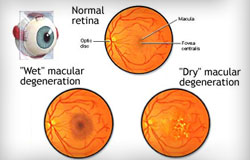
    Age-related macular degeneration (AMD) is an eye disorder which usually affects older adults and results in a loss of vision in the center of the visual field (the macula) because of damage to the retina. It occurs in “wet” and “dry” forms. It is a major cause of blindness and visual impairment in adults older than 50 years old. People with macular degeneration often find it hard to recognize faces, read, and it can significantly affect their quality of life.

    In the dry form of AMD, cellular debris accumulates in the macula (central part of the retina) and this is called drusen. In the wet form of AMD, new blood vessels grow in the layers of the macula and they bleed easily. However, macular degeneration will not lead to total blindness. In almost all cases, some vision remains. Approximately 10% of patients 66 to 74 years of age will have findings of macular degeneration. The prevalence increases to 30% in patients 75 to 85 years of age.

    Risks for macular degeneration include high blood pressure, smoking, high cholesterol, obesity, consumption of large amounts of polyunsaturated fats, and Caucasian race. The lifetime risk of developing late-stage AMD is 50% for people who have a relative with AMD, versus 12% for people who do not have relatives with AMD.

    Macular Degeneration Treatment for the dry form of AMD involves ultraviolet light protection, smoking cessation, diet modification, and an increased intake of two carotenoids, lutein and zeaxanthin. A good AMD Prevention Diet includes dark green vegetables, brightly colored fruits and vegetables, no simple sugars, cold water fish such as salmon, mackerel and sardines, and well as berries. Using olive oil or nut oil, as opposed to partially hydrogenated vegetable oil, is also beneficial. Consuming omega-3-fatty acids has been correlated with a reduced progression of AMD, also in conjunction with a low glycemic index diet. The Age-Related Eye Disease Study in 2001 showed that consumption of vitamins with high levels of vitamin C, vitamin E, beta-carotene, zinc and copper could help to delay the progression of dry AMD.

    Treatment of the wet form of AMD uses injectable drugs that cause regression of the abnormal blood vessels in the macula. These drugs, called antiangiogenics or anti-vascular endothelial growth factors, are injected into the vitreous, which is the jelly-like portion in the back of the eye.

  • What is Dry Eye Disease?

    To keep your eyes comfortable and your vision optimal, a normal, thin film of tears coats your eyes. Three main layers make up this tear film:

    1. The innermost layer is the Mucin. This layer is produced by the cells in the conjunctiva, which is the clear layer with blood vessels, over the white portion of the eye. The mucin layer helps the tear film spread evenly over the eye.
    2. The middle, or aqueous layer, is the largest and thickest. This layer is a very dilute saltwater solution. The lacrimal glands under the upper eyelid produce this watery layer. This layer gives moisture to the eye and flushes out any dust, debris or allergens that may get into the eye.
    3. The most superficial layer is a very thin layer of lipids (fats or oils). These lipids are produced by the meibomian glands (oil glands in the eyelids). The main function of this lipid layer is the help decrease evaporation of the watery layer beneath it.

    Dry Eye Syndrome (DES) is a very common eye disorder that results from a deficiency in one or more layers of the tear film. The estimated number of people affected by DES in the United States ranges from 25-30 million. Dry Eye Syndrome is more common in women than men. Causes of dry eye includes conditions that cause decreased tear production, excessive tear evaporation or an abnormality in the production of the lipids in the tear film. Some causes include hormonal changes, autoimmune diseases such as Sjogren’s Syndrome, rheumatoid arthritis or lupus. Some medications, such as antihistamines, antidepressants, blood pressure medications, and oral contraceptives, may decrease tear production. Insufficient lipid layers are the result of the dysfunction of the Meibomian glands, which is common in rosacea and blepharitis (inflammation of the eyelids).

    Symptoms of DES include dry, gritty sensation, or filmy feeling in the eyes. There also may be burning or itching, redness, blurred vision, foreign body sensation and light sensitivity. Sometimes a symptom of Dry Eye Syndrome may actually be intermittent excessive tearing. When the eye becomes irritated, it may initiate reflex tearing with production of a large amount of tears all at once to try to get moist and comfortable again.

    Treatment of Dry Eye Syndrome is multi-tiered. Tips at home include: a humidifier to put more moisture in the air, decrease the use of ceiling fans, and changing the air filter in your home frequently to remove irritating allergens. Other initial treatment options include artificial tears and lubricating eyedrops and gels, as well as lubricating ointments at night while you are asleep. Although no cure for DES exists, many medical treatments are now available. Cyclosporine A 0.5% (Restasis®) helps to decrease the inflammation in the lacrimal gland and on the ocular surface, which allows the eye to produce healthier tears. Corticosteroid drops may be used either alone or with Restasis®, although the use of these drugs must be monitored by your ophthalmologist since side effects may occur. Nonsteroidal anti-inflammatory drops, antibiotic drops and pills, and serum tears from the patient’s own blood also have a role in the treatment of DES.

    Dr. Cottle now has a specific Dry Eye Clinic at Lake Austin Eye and offers even more testing and treatment options, including Tear Osmolarity, Sjogren’s Syndrome early detection, and LipiFlow treatment for lid diseases. Please refer to the section titled Dry Eye Clinic.

  • What is Blepharitis?

    Blepharitis is an eye disorder that is caused caused by inflammation of the eyelids. Blepharitis occurs in two forms, anterior and posterior. Anterior blepharitis affects the outside front of the eyelid, where the eyelashes are attached. The two most common causes of anterior blepharitis are bacteria (Staphylococcus) and scalp dandruff. Posterior blepharitis affects the inner eyelid and is caused by problems with the oil glands in the eyelid (Meibomian glands). Two skin disorders can cause this form of blepharitis: rosacea and seborrheic dermatitits. When posterior blepharitis becomes severe, a blockage of the gland may occur, which is called a chalazion.

    Treatment of blepharitis includes lid scrubs with a dilute soapy solution, as well as warm lid compresses to soften the lipid contents of the eyelid glands. Various medications such as antibiotic drops, corticosteroid drops and oral antibiotics may be prescribed to control blepharitis.

  • How does Diabetes affect the Eye?

    Diabetes causes changes in the blood vessels in the retina, the back layer of the eye that contains nerve cells responsible for transmitting light images to the brain. Diabetes makes the blood vessels balloon and form pouches, which makes them faulty and leaky. This can severely affect a person’s vision, especially if the leaking fluid occurs in the macula, the center part of the retina responsible for detail vision. If the damage to the blood vessels continues untreated, the vessels then close off and new, weaker blood vessels grow back in these areas. Bleeding occurs and scar tissue forms, which can lead to retinal detachment and further loss of vision.

    Your retina can be badly damaged before you notice any change in vision. Several factors can increase your risk of developing diabetic retinopathy: poor blood sugar control, high blood pressure levels, amount of time you have had diabetes, and genetic factors. A person with diabetes should have a complete dilated exam once a year to determine if there are any signs of retinopathy.

  • What is Fuch’s Corneal Dystrophy?

    Fuch’s Dystrophy is an inherited disease of the cornea in which the inner most layer of the cornea, called the endothelium, becomes dysfunctional and may result in decreased vision.  The front part of a normal eye is filled with fluid called the aqueous.  However, the cornea, which separates the outside air from the aqueous fluid inside the eye, must maintain a proper balance of moisture in order to maintain its clarity.  To accomplish this task, the inside of the cornea is lined with a specialized layer of cells called the endothelium.  The endothelial cells function as one-way pumps, drawing fluid out of the cornea and pumping it back inside the eye.  The cornea, despite being in direct contact with the aqueous eye fluid, doesn’t become overly hydrated and maintains clarity.

    In Fuch’s Dystrophy, the endothelial cells are slowly replaced with small collagen bumps called guttata.  Guttata may first appear in the cornea in the 20’s and 30’s, though rarely cause any symptoms until later in life.  As a person ages, the guttata may slowly increase in number and size.  In the process, the endothelial cells surrounding them may slowly die.  When a critical number of endothelial cells are lost, usually by the age of 50 or 60 years, the pump function of the endothelium may be overwhelmed and the cornea starts to swell with eye fluid.  As the swelling worsens, the vision may start to decrease.  In cases where large amounts of guttata exist on the cornea, the vision may also become compromised even without corneal swelling, due to irregular scattering of light passing through the dense guttata layer.

    Fuch’s Dystrophy is caused by an inherent malfunction in the way the inner lining of cells behave.  The gene is transferred from parent to child in an autosomal dominant fashion, which means that each child of an affected parent has a 50% chance of developing the disease.  Though many family members may have Fuch’s Dystrophy, its effects may vary greatly from one family member to the next.  As such, some individuals with Fuch’s Dystrophy will have signs of the eye disease visible only to a doctor examining their corneas with a slit lamp, and will never suffer any visual loss from the disorder.  Other individuals who have worse forms of the disease may suffer slowly progressive visual loss.

  • What is Keratoconus?

    Keratoconus literally means “cone-shaped cornea”. The normal cornea surface is smooth and dome shaped and aspheric (flattens towards the edges). Keratoconus is an eye disorder characterized by an irregular corneal surface (cone-shaped) resulting in blurred and distorted images. The condition is often progressive, and results in increasing levels of nearsightedness and astigmatism that have to be corrected with special glasses or gas-permeable contact lenses. Since the cornea is responsible for refracting most of the light entering the eye, corneal abnormalities can result in significant visual impairment, making simple tasks difficult like driving or reading.

    Thankfully, Lake Austin Eye offers Corneal Cross-Linking, designed to halt the progression of Keratoconus, and covered by most insurance carriers.

    Symptoms of keratoconus include mild burning, glare at night, irritable eye, sensitivity to light, and distortion of vision. According to the American Academy of Ophthalmology, about 1/2000 people will develop keratoconus. Most people will have a mild or moderate form of the disease. Less than 10% of people with keratoconus will develop the most severe form. It typically is diagnosed in the late teens or twenties, however, many people have been diagnosed in their mid to late thirties. It is common for one eye to progress faster than the other and the eyes may go for long periods of time without any change, and then change dramatically over a period of months. Rubbing the eyes is known to hasten progression, and modern treatments such as Intacs and collagen cross linking may delay progression.

    CLICK HERE to Request an In-Office Examination and Consultation
    (Most Major Insurance Carriers Accepted!)

  • What is Conjunctivochalasis?

    The white of the eye, or the sclera, is covered by a layer of skin called the conjunctiva. This layer of skin can become redundant and bunch up in the corners of the eyes, which is called Conjunctivochalasis. Conjunctivochalasis can become symptomatic, leading to sensation of dry eyes [link to dry eye page/paragraph], redness, eye fatigue, unstable tear film, foreign body sensation, or a constant “presence” of the eyes. If it is severe, it can even cause blurred vision, discharge, and recurrent conjunctival bleeding.

    A direct cause for conjunctivochalasis remains unknown, however research suggests that this may occur due to impaired lymphatic flow, mechanical trauma, or UV light damage. Treatment may include artificial tears, however if severe, fornix reconstruction surgery (removal of the redundant conjunctival tissue) may be required to restore the normal tear reservoir.